Uterine fibroids are non-cancerous growths that develop in or on the uterus. They are common among women of reproductive age. Detecting fibroids is crucial, especially for women experiencing symptoms like heavy periods or infertility. Ultrasound is a primary tool used to detect these growths. This article explores how effective ultrasounds are in identifying fibroids and their role in managing female infertility.
Understanding Uterine Fibroids
Fibroids, also known as leiomyomas or myomas, are composed of muscle and fibrous tissue. They vary in size and can be as small as a seed or as large as a melon. Depending on their location, fibroids are classified into:
Intramural Fibroids: Found within the muscular wall of the uterus.
Submucosal Fibroids: Protrude into the uterine cavity.
Subserosal Fibroids: Extend to the outside of the uterus.
While many women with fibroids experience no symptoms, others may face challenges like heavy menstrual bleeding, pelvic pain, or fertility issues.
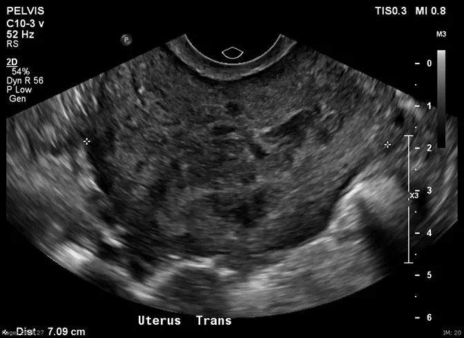
The Role of Ultrasound in Detecting Fibroids
Ultrasound imaging uses sound waves to create pictures of the inside of the body. It’s a non-invasive method commonly used to detect uterine fibroids. There are two main types of pelvic ultrasounds:
– **Transabdominal Ultrasound**: The probe is moved over the abdomen.
– **Transvaginal Ultrasound**: The probe is inserted into the vagina, providing a closer view of the uterus.
Transvaginal ultrasounds are generally more effective in detecting smaller fibroids due to their proximity to the uterus.
Effectiveness of Ultrasound in Fibroid Detection
Ultrasounds are effective in identifying most fibroids, especially larger ones. However, their effectiveness can vary based on several factors:
– **Size of the Fibroid**: Larger fibroids are easier to detect. Very small fibroids might be missed.
– **Location**: Fibroids located at the back of the uterus or deep within the uterine wall can be harder to visualize.
– **Number of Fibroids**: Multiple fibroids can sometimes overlap, making individual detection challenging.
– **Technician’s Expertise**: The skill of the person performing the ultrasound plays a significant role in accurate detection.
– **Quality of Equipment**: Advanced ultrasound machines provide clearer images, aiding in better detection.
Limitations of Ultrasound
While ultrasounds are a valuable diagnostic tool, they have limitations:
– **Small Fibroids**: Fibroids smaller than 1 cm might not be detected.
– **Obesity**: Excess body fat can interfere with image clarity.
– **Bowel Gas**: Gas in the intestines can obstruct the view of the uterus.
– **Similar Appearance to Other Conditions**: Some fibroids may resemble other uterine abnormalities, making diagnosis challenging.
In such cases, additional imaging tests like MRI might be recommended for a more detailed view.
Advanced Ultrasound Techniques
To enhance fibroid detection, several advanced ultrasound techniques are employed:
– **3D Ultrasound**: Provides a three-dimensional view of the uterus, helping in accurate localization of fibroids.
– **Saline Infusion Sonography (SIS)**: Involves injecting saline into the uterine cavity during ultrasound to get a clearer view of the uterine lining and detect submucosal fibroids.
– **Color Doppler Ultrasound**: Assesses blood flow within fibroids, helping differentiate them from other masses.
These techniques improve the accuracy of fibroid detection and aid in treatment planning.
Fibroids and Female Infertility
Fibroids can impact fertility, depending on their size and location. Submucosal fibroids, which protrude into the uterine cavity, are most likely to interfere with implantation and pregnancy. Intramural fibroids can also affect fertility if they distort the uterine cavity.
For more information on how fibroids relate to infertility, you can visit the following resources:
– Female Infertility Types
– Female Infertility Symptoms
– Female Infertility Treatment
Treatment Options Based on Ultrasound Findings
Once fibroids are detected via ultrasound, treatment options vary based on symptoms, size, and location:
– **Watchful Waiting**: If fibroids are small and asymptomatic, regular monitoring might be sufficient.
– **Medications**: Hormonal treatments can help shrink fibroids and alleviate symptoms.
– **Minimally Invasive Procedures**: Techniques like uterine artery embolization cut off blood supply to fibroids, causing them to shrink.
– **Surgical Options**:
– **Myomectomy**: Surgical removal of fibroids, preserving the uterus.
– **Hysterectomy**: Complete removal of the uterus, recommended in severe cases.
The choice of treatment depends on individual circumstances and reproductive goals.
Importance of Regular Monitoring
Regular ultrasounds are essential for women with known fibroids, especially if they plan to conceive. Monitoring helps:
– Track fibroid growth.
– Assess the effectiveness of treatments.
– Make timely decisions about interventions.
Early detection and management can prevent complications and improve fertility outcomes.
Conclusion
Ultrasound is a vital tool in detecting uterine fibroids, offering a non-invasive and effective method for diagnosis. While it has limitations, especially in detecting very small or deeply located fibroids, advancements in ultrasound technology have enhanced its accuracy. Understanding the role of fibroids in female infertility underscores the importance of regular monitoring and timely intervention. By staying informed and proactive, women can make informed decisions about their reproductive health.
Related topics:


