Cervical polyps are small, benign growths that develop on the cervix. While they are often harmless, they can sometimes cause symptoms like abnormal bleeding or discharge. Detecting these polyps is crucial for proper management and to rule out more serious conditions. One of the primary methods for identifying cervical polyps is through ultrasound imaging. This article explores how cervical polyps appear on ultrasounds, the techniques used for detection, and the implications for patient care.
Understanding Cervical Polyps
Cervical polyps are protrusions that arise from the mucosal surface of the cervix. They are typically less than 3 cm in diameter and can be either pedunculated (attached by a stalk) or sessile (broad-based). Most cervical polyps are asymptomatic and are discovered incidentally during routine pelvic examinations. However, some women may experience symptoms such as:
– Intermenstrual bleeding
– Postcoital bleeding
– Abnormal vaginal discharge
While the exact cause of cervical polyps is not well understood, factors like chronic inflammation, hormonal imbalances, and vascular congestion are believed to contribute to their development.
The Role of Ultrasound in Detecting Cervical Polyps
Ultrasound imaging is a non-invasive diagnostic tool that uses sound waves to create images of internal body structures. In gynecology, transvaginal ultrasound (TVUS) is commonly used to evaluate the uterus and cervix. Cervical polyps can often be visualized during a TVUS, appearing as well-defined masses within the endocervical canal.
Ultrasound Features of Cervical Polyps
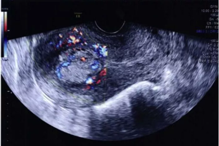
On ultrasound, cervical polyps typically present as:
– Well-circumscribed, echogenic or hypoechoic masses within the endocervical canal
– Pedunculated or sessile structures
– Presence of a feeding vessel, identifiable using color Doppler imaging
Identifying the stalk or feeding vessel is crucial, as it helps differentiate cervical polyps from other intrauterine or endometrial lesions.
Advanced Ultrasound Techniques
In some cases, standard transvaginal ultrasound may not provide sufficient detail to accurately identify cervical polyps. Advanced imaging techniques can enhance visualization:
– **Color Doppler Ultrasound**: This technique assesses blood flow within the polyp, helping to identify the feeding vessel.
– **3D Ultrasound**: Provides a more detailed, three-dimensional view of the cervix and any present polyps.
– **Sonovaginography**: Involves the introduction of ultrasound gel into the vagina to improve the acoustic window, enhancing the visualization of cervical structures.
These advanced techniques can improve diagnostic accuracy, especially in complex cases or when polyps are not clearly visible on standard ultrasound.
Clinical Significance of Detecting Cervical Polyps
While most cervical polyps are benign, their detection is important for several reasons:
– **Symptom Management**: Identifying the polyp can explain symptoms like abnormal bleeding, allowing for appropriate treatment.
– **Exclusion of Malignancy**: Although rare, some cervical polyps can harbor precancerous or cancerous cells. Detection allows for histological examination to rule out malignancy.
– **Fertility Considerations**: Large polyps can obstruct the cervical canal, potentially impacting fertility. Their removal can alleviate this issue.
Management of Cervical Polyps
The treatment approach for cervical polyps depends on factors like size, symptoms, and patient age:
– **Observation**: Small, asymptomatic polyps may not require immediate intervention and can be monitored over time.
– **Polypectomy**: Symptomatic or large polyps are typically removed surgically. This can often be done in an outpatient setting using forceps or electrosurgical techniques.
– **Histological Examination**: Removed polyps should be sent for pathological analysis to confirm their benign nature and rule out malignancy.
Post-removal, patients are usually advised to undergo follow-up examinations to monitor for recurrence.
Conclusion
Cervical polyps are common, usually benign growths that can be effectively detected using ultrasound imaging. Transvaginal ultrasound, especially when combined with advanced techniques like color Doppler and 3D imaging, provides a reliable method for identifying these lesions. Early detection is key to managing symptoms, ensuring proper treatment, and ruling out more serious conditions. Regular gynecological check-ups and imaging studies play a vital role in maintaining cervical health.
Related topics:


